-
Products
- Gas analysis systems
- GAOS SENSON gas analyzers
- GAOS MS process mass spectrometry
- MaOS HiSpec ion mobility spectrometer
- MaOS AxiSpec ion mobility spectrometer
- Applications
- News
- Events
- About us
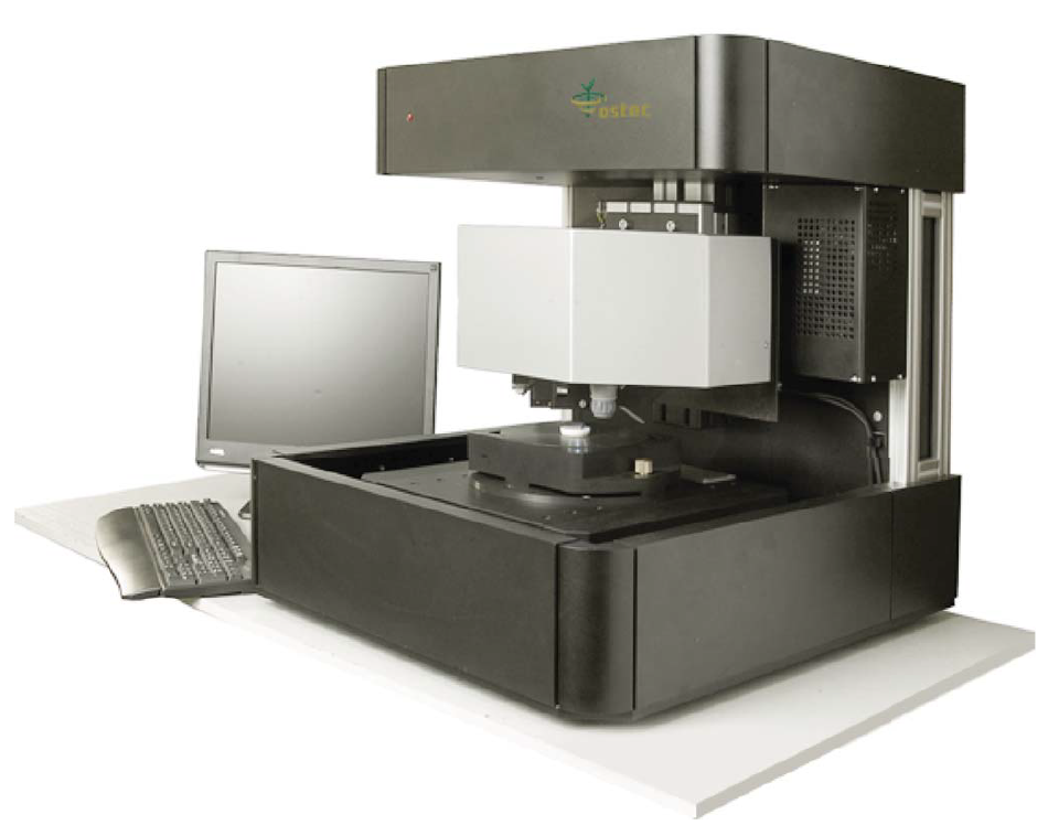
XROS MF30 – laboratory x-ray microscope-microprobe for studies of the objects by the methods of the optical microscopy, radiography, local element XRF microanalysis with the possibility of the element mapping.
Using a microscope, a sample of up to 400 mm in size along the Y-axis and of unlimited size along the X-axis (max. scan area 150×150 mm; in the case of a larger area, the scanned areas can be stitched) and up to 105 mm high can be performed.
An overview video camera and two optical microscopes with magnification up to 200 times are using for accurate determination of the scanning area.
The central optical microscope with automated sharpness adjustment is combined with the axis of the microprobe (axis of the x-ray beam).
Local X-ray fluorescence microanalysis with the possibility of elemental mapping and X-ray studies can be carried out both separately and simultaneously.
The sample positioning accuracy is 10 microns.
The minimum diameter of the x-ray probe is 30 µm.
The range of simultaneously measured elements from 11Na to 92U.
Samples: fragments of targets and textiles with bullet holes.
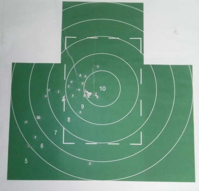
The target from a shooting gallery after shooting from 10 and 15 meters distances was investigated (Figure 1). Small pieces of the target with traces of a shot were selected. Elemental mapping of selected target areas was performed.
 |
 |
|
Figure 1. Shooting target with bullet holes |
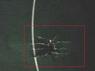
Figure 2. An optical image from a built-in video camera with the highlighted scan area |
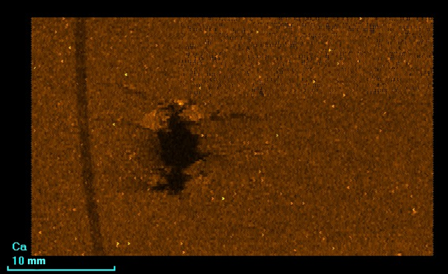
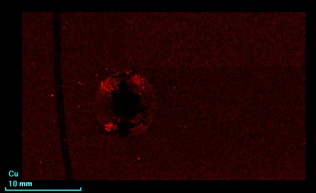
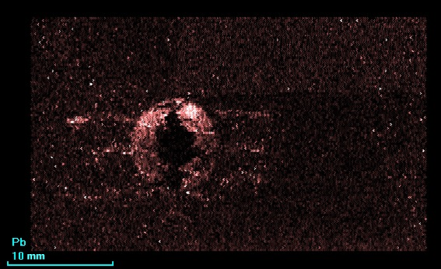
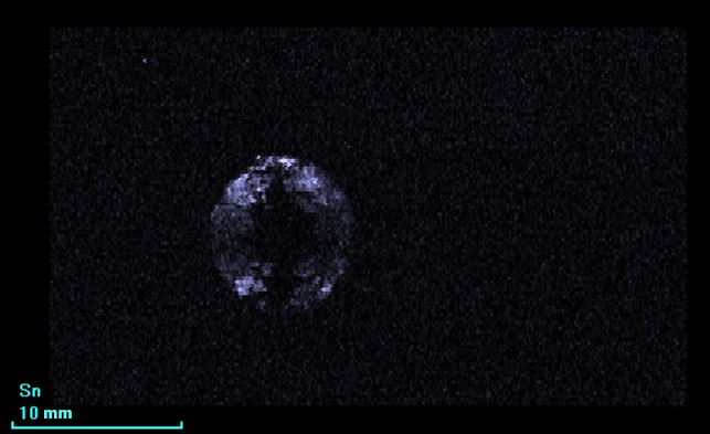
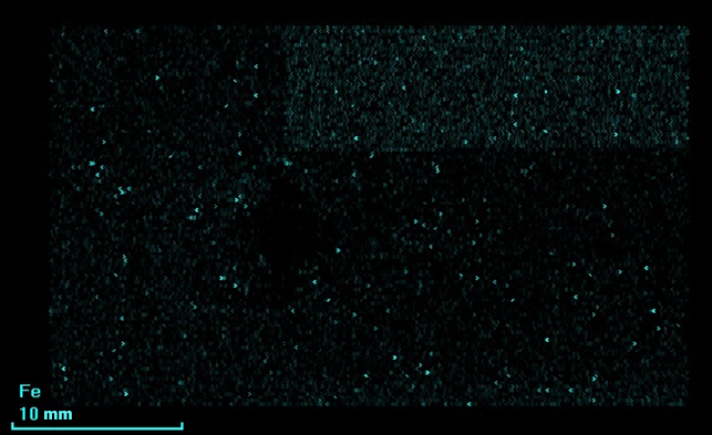
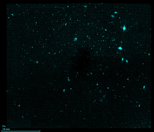
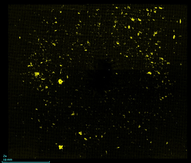
The following are the results of the elemental mapping of the scan area from Figure 2.
 |
 |
 |
 |
 |
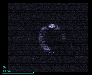
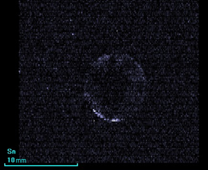
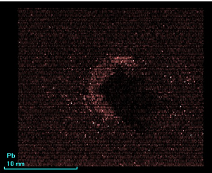
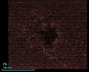
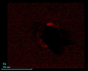
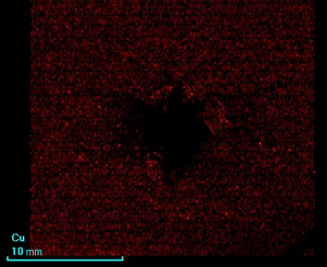
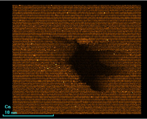
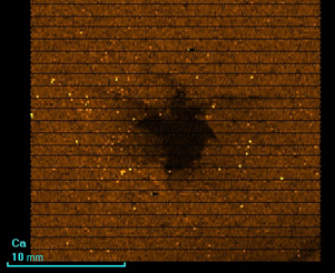
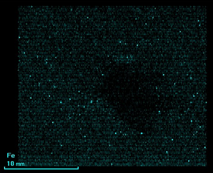
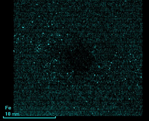
The following are the comparison of the results of targets elemental mapping (on the left – 10 m shooting distance, on the right – 15 m shooting distance).
|
|
|
|
|
|
|
|
|
|
 |
 |
 |
 |
 |
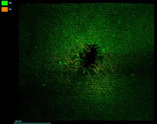 |
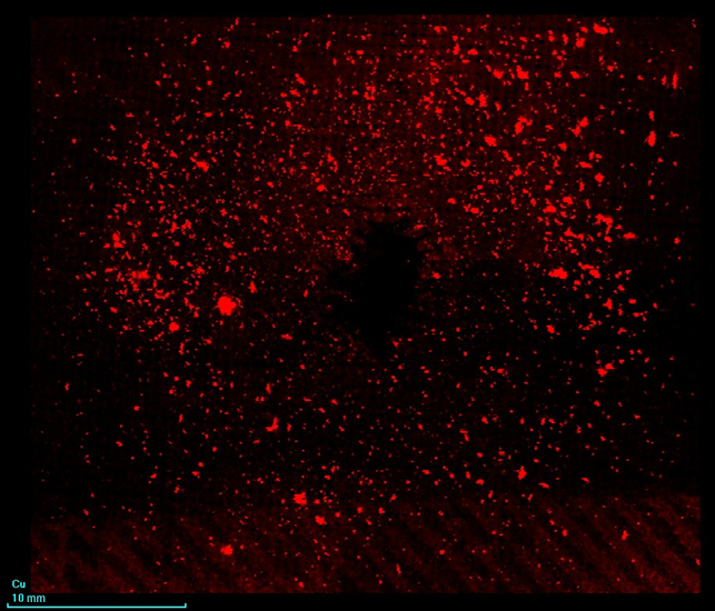
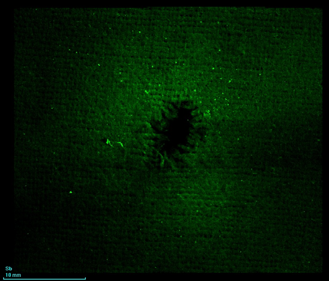
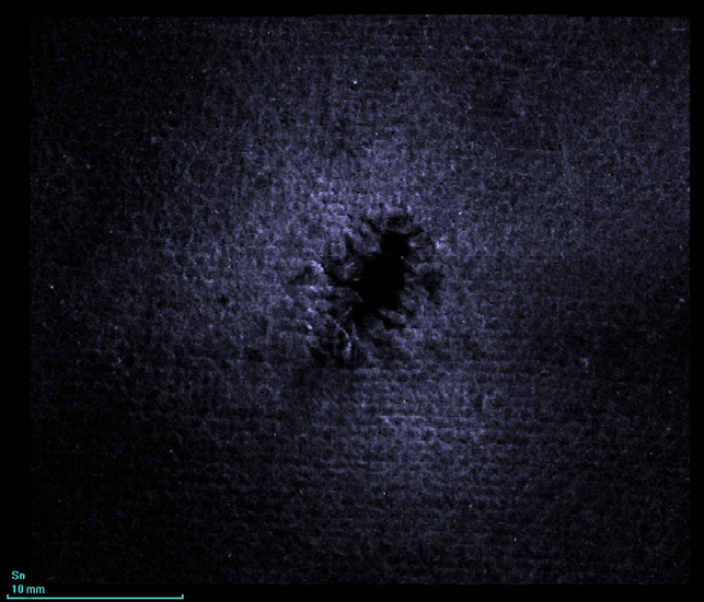
As a result of the investigation the presence on the target material (paper and textile) of microinclusions of such elements as Mn, Fe, Ni, Cu, Zn, Pb was detected. The density of these points decreases with distance from the bullet hole, It means that they are products of the shot.
Using the built-in software of the mathematical methods, the size of points with microinclusions was estimated. The maximum diameter of microinclusions is estimated to be about 40 microns. Most microinclusions have a smaller diameter.
XROS MF30 x-ray analytical microscope-microprobe allows analysis of shot residual with a high spatial resolution and elemental sensitivity.
|
Scan step 200 µm |
Electric current 10 000 µA |
|
Scan rate 200 µm/s |
XRT Mo anode |
|
Measurement time 500 ms |
Atmosphere Air |
|
Voltage 30 kV |
|