RAMOS N500
3D Scanning Laser Raman Microscope

Simultaneous / Multifunctional Analysis
-
Raman Measurements
-
Luminescence Measurements
-
Laser Reflection & Transmission Measurements
-
Spectral and Polarization measurements

3D high-contrast images in reflected light
3D confocal Raman measurements
Confocal Detection Principle
Confocal Laser Scanning Raman Microscope has become a widely recognized research instrument in recent years. Confocal microscopy offers several advantages over conventional wide-field optical microscopy, including the ability to control depth of field, elimination or reduction of background information away from the focal plane and the capability to collect serial optical sections from thick samples. The image of the extended sample is generated by scanning the focused laser beam across a defined area.
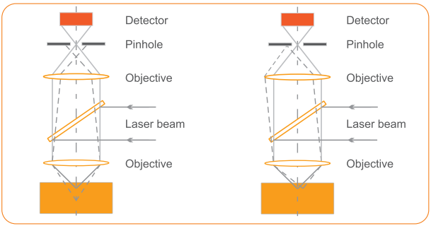
The pinhole aperture rejects the residual scattered rays originated from any out-of-focus points on a sample.
We have created the instrument that is right for you

High spectral resolution

Spatial resolution: less than 500 nm (Z), 200 nm (X, Y)
Spectral resolution: ~ 0.25 cm-1
Wavelength accuracy in spectrum with CCD detector: 0.005 nm (1800 l / mm)
Applications
Semiconductors
High spatial resolution Raman confocal microscopy can provide information on dopant concentrations and stress distribution in semiconductor materials.
Biology
Raman spectroscopy allows easy visualization of cellular components with minimum perturbation.
Pharmaceutics
Confocal Raman spectroscopy allows chemical compounds and molecular conformers in various drugs to be identified and their distribution mapped with high spatial resolution.
Geology
Confocal Raman microscopy is an excellent technique for characterization of minerals, detection of components distribution and their phase transitions.
Cosmetology
Confocal Raman microspectroscopy is a promising technique which enables measuring the skin care products as well as their penetration capability.
Forensics
Application areas include identification of unknown substances, different types of fibers, glasses, paints, explosive materials, inks, narcotic and toxic substances, proof of authenticity of documents.
Material science
Confocal Raman offers excellent spatial resolution for characterization of materials (superconductor, polymers, coatings, composites, carbon nanotubes, graphene, etc.).
Heritage and Art, Gemology
Raman spectroscopy allows identfication of pigments and binders used in paintings. The spectroscopic analysis of archaeological samples (ceramics, glasses, etc.) provides information on their origin and history. Raman technique allows rapid identfication of colored stones, natural and synthetic diamonds.
and many more…
Raman megapixel image for 3 sec
Fully automated system with up to 5 integrated lasers

High spatial resolution and sensitivity
Major features
The highest spectral and imaging resolution with specially designed spectrometer
Specially designed imaging spectrometer incorporates many features that make it ideal for confocal Raman measurements. The image of pinhole is projected to a multichannel detector without any aberrations.
The smaller amount of illuminated pixels on the CCD matrix leads to the smaller dark counts and the higher spectral resolution.
Spectral resolution of RAMOS N500 with an Echelle grating is 0.25 cm
-1.

Spectral image of the pinhole on the CCD camera (aberration free).
CCD pixel size is 12 μm.
High optical throughput for enhanced sensitivity
The 4th order Silicon band at 1940 cm
-1 can be observed in less than one minute using a low intensity laser.
2D / 3D images can be acquired rapidly.

Silicon 4th order sensitivity.
Fully automated
People with little or no experience in Raman spectroscopy can use RAMOS N500. The system is highly modular and fully automated. Up to 5 lasers can be used.
The lasers can be switched from one to another by just one click.
Motorized control for laser power, beam diameter, polarization orientation, pinhole size and grating is provided.

Low frequency Raman shift measurements (down to 5 cm-1) with Bragg Super-Notch filters
True confocal design
High spatial resolution
Laser Raman microscope RAMOS N500 can achieve:
- lateral resolution close to theoretical limitation
Laser
wavelength, nm
|
Objective
|
XY - plane
resolution, nm
|
|
488
|
100x, NA = 0.9
|
250
|
|
532
|
100x, NA = 0.9
|
275
|
|
633
|
100x, NA = 0.9
|
320
|
|
785
|
100x, NA = 0.9
|
390
|

RAMOS N500 can take high definition Raman images (λ = 514 nm, 100x, NA = 1.4).
- axial resolution (in depth direction, 100x, NA = 0.9)
Laser wavelength, nm
|
Z (axial) resolution, nm
|
|
488
|
520
|
|
532
|
560
|
|
633
|
660
|
|
785
|
800
|

Axial resolution of 450 nm (λ = 488 nm, 100x, NA = 0.95).
Wide Raman shift measurement range
Laser wavelength, nm
|
Wavenumber range, cm-1
|
|
325
|
125 - 8000
|
|
355
|
115 - 8000
|
|
473
|
80 - 6000
|
|
532
|
50 - 8000
|
|
633
|
50 - 6000
|
|
785
|
40 - 2800
|
Low-frequency Raman shift measurement range can be expanded using Bragg notch filters.

Low-frequency Raman bands of sulfur (lower than 250 cm-1, 633 nm laser)
Megapixel Raman image for 3 sec
True confocal design
High spatial resolution
3D scanning laser confocal Raman microscope RAMOS N500 provides the acquisition of two images within a single scan: a Rayleigh image (using laser light reflected from a sample) and a spectral image by Raman scattering.
Ultrafast imaging option allows to get confocal image in 3 sec (3 μs/pixel).
RAMOS N500 uses fast beam scanning by galvano mirrors.
Layout of galvano mirror scanner module allows mapping with no intensity losses from image center to its edges.


Rayleigh (1000 x 1000 pixels, time per 1 pixel is 3 μs) and Raman (1000 x 1000 pixels, time per 1 pixel is 43 μs) images of Granite Gneiss India.
Anatase distribution.
Fast imaging mode with EMCCD / CCD
RAMOS N500 system can be used with a number of different detectors.
Up to three detectors can be used simultaneously. Proprietary algorithm for taking high speed of Raman imaging with fast spectral CCD (EMCCD) is offered.
The use of an EMCCD (Electron Multiplying CCD) camera can greatly increase Raman detection efficiency and speed.

Raman image of Silicon / SiO2 sample.
Si distribution (500 x 500 pixels, time per pixel is 5 ms).
Fully automated system Software package with powerful analytical functionality
Ultrawide field Raman imaging
Uniform, large size scanning area of a galvanic scanner module:
- 150 μm x 150 μm (objective lens 100x)
- 320 μm x 320 μm (objective lens 40x)
- 680 μm x 680 μm (objective lens 20x)
Automatic XY stage can be used for ultra-wide field imaging.
The panoramic image (hyper image) by automatic stitching of a series of images obtained with the use of galvanic scanner.

High precision spectrometer calibration
RAMOS N500 is equipped with a neon lamp (option) for spectral calibration.
Calibration is possible at any wavelength by one click in the control software.
More capabilities
- microscope can be equipped with a heating or cooling stage, vacuum or high pressure cell
- fiber optics probe for remote measurements

Data Acquisition and Data Analysis software
RAMOS N500 software “Nano SPO” with powerful analytical functionality is designed for hardware operating, data acquisition and data analysis.
- 2D and 3D image creation
- Autofocus control during mapping
- Automatic background subtraction, cosmic ray removing, peak shift imaging, etc.
- Support for external spectral databases
- Data export to popular file formats
- Intuitive user-friendly interface
- Compatible with Windows XP, Vista, 7


